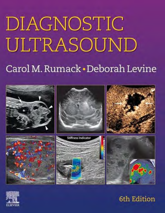
It is an honor to present to you the sixth edition of Diagnostic
Ultrasound. As in previous editions, we have included experts
in the field to share their authoritative experience. Given the
nature of ultrasound practice, which in radiology departments
is moving toward a more body imaging/organ-based approach,
this edition has been expanded to include more correlative
imaging, and thus is a substantial update from the previous
versions. We have also greatly expanded our video library to
enable real-time examples of anatomy and pathology.
Previous editions have been very well accepted as reference
textbooks and have been the commonly used reference in ultrasound education and practices worldwide. The text and
references have all been updated and are available online. We
are pleased to provide over 2,300 images, with 657 videos of
ultrasound anatomy and pathology that complement the static
images in the book. The display of real-time ultrasound has
helped to capture those areas of anatomy that are difficult to
depict on a still image. The video sweeps also allow a more
complete depiction of pathology, and the relationship of pathology to surrounding anatomy.
Another new feature of this book is incorporating artifacts
into the physics chapter, which allows the reader to better
appreciate the relationship between the creation of images and
the artifacts that help define intrinsic details of tissue.
41 outstanding new and 43 continuing authors have contributed to this edition, and all are recognized experts in the
field of ultrasound. As in previous editions, we have emphasized the use of collages to show many examples of similar
anatomy and pathology. These images reflect the spectrum of
sonographic changes that may occur in a given disease, instead
of only the most common manifestation.
We have again used colored boxes to highlight the important or critical features of sonographic diagnoses. Key terms
and concepts are emphasized in boldface type. To direct the
readers to other research and literature of interest, comprehensive updated reference lists are provided.
Diagnostic Ultrasound is again divided into two volumes.
Volume I consists of Parts I to III. Part I contains chapters on
physics (including artifacts and elastography) and biologic effects of ultrasound. It also includes description of ultrasound
contrast technique and images, with an entirely new section on
starting an ultrasound contrast practice in your laboratory. Part
II covers abdominal, pelvic, retroperitoneal, interventional,
and hernia sonography. Part III covers vascular sonography and
small parts imaging, including testicular, breast, thyroid, and
a newly expanded chapter on parathyroid and other glands in
the head and neck. Volume 2 comprises Part IV (gynecology),
Part V (obstetrics), and Part VI (pediatrics). Each of these sections has been expanded to include correlative imaging and
description of the value of elastography and ultrasound contrast
when appropriate.
Diagnostic Ultrasound is for practicing physicians, residents,
medical students, sonographers, and others interested in understanding the vast applications of diagnostic and interventional sonography in patient care. Our goal is for Diagnostic
Ultrasound to continue to be the most comprehensive reference
book available in the sonographic literature, with a highly
readable style and superb images and video clips.
Deborah Levine, MD, FACR
Carol M. Rumack, MD, FACR
Trân trọng giới thiệu tới các bạn ấn bản thứ sáu của cuốn sách Chẩn đoán
Siêu âm. Như trong các phiên bản trước, chúng tôi đã bao gồm các chuyên gia
trong lĩnh vực này để chia sẻ kinh nghiệm có thẩm quyền của họ. Cho
bản chất của thực hành siêu âm, trong khoa X quang
đang hướng tới một cách tiếp cận dựa trên hình ảnh cơ thể/cơ quan nhiều hơn,
ấn bản này đã được mở rộng để bao gồm nhiều thông tin tương quan hơn
hình ảnh, và do đó là một bản cập nhật đáng kể từ phiên bản trước
các phiên bản. Chúng tôi cũng đã mở rộng đáng kể thư viện video của mình sang
cho phép các ví dụ thời gian thực về giải phẫu và bệnh lý.
Các phiên bản trước đã được chấp nhận rất tốt làm tài liệu tham khảo
sách giáo khoa và là tài liệu tham khảo thường được sử dụng trong giáo dục và thực hành siêu âm trên toàn thế giới. Văn bản và
tài liệu tham khảo đều đã được cập nhật và có sẵn trực tuyến. Chúng tôi
hân hạnh cung cấp hơn 2.300 hình ảnh, với 657 video về
siêu âm giải phẫu và bệnh lý bổ sung cho tĩnh
hình ảnh trong sách. Màn hình hiển thị siêu âm thời gian thực có
đã giúp nắm bắt được những khu vực giải phẫu khó
khắc họa trên ảnh tĩnh. Việc quét video cũng cho phép nhiều hơn
mô tả đầy đủ về bệnh lý và mối quan hệ của bệnh lý với giải phẫu xung quanh.
Một điểm mới khác của cuốn sách này là kết hợp các hiện vật
vào chương vật lý, cho phép người đọc hiểu rõ hơn
đánh giá cao mối quan hệ giữa việc tạo ra hình ảnh và
các tạo tác giúp xác định các chi tiết bên trong của mô.
41 tác giả mới xuất sắc và 43 tác giả tiếp tục đóng góp cho ấn bản này và tất cả đều là những chuyên gia được công nhận trong lĩnh vực này.
lĩnh vực siêu âm. Như trong các phiên bản trước, chúng tôi đã nhấn mạnh việc sử dụng ảnh ghép để hiển thị nhiều ví dụ về các tác phẩm tương tự.
giải phẫu và bệnh lý. Những hình ảnh này phản ánh quang phổ của
những thay đổi siêu âm có thể xảy ra trong một bệnh nhất định, thay vào đó
chỉ là biểu hiện phổ biến nhất.
Chúng tôi lại sử dụng các hộp màu để làm nổi bật những đặc điểm quan trọng hoặc then chốt của chẩn đoán siêu âm. Điều khoản quan trọng
và các khái niệm được nhấn mạnh bằng kiểu in đậm. Để chỉ đạo
độc giả đến các nghiên cứu và tài liệu quan tâm khác, danh sách tài liệu tham khảo cập nhật toàn diện được cung cấp.
Siêu âm chẩn đoán một lần nữa được chia thành hai tập.
Tập I gồm có Phần I đến Phần III. Phần I gồm các chương về
vật lý (bao gồm cả tạo tác và đo độ đàn hồi) và tác dụng sinh học của siêu âm. Nó cũng bao gồm mô tả về siêu âm
kỹ thuật và hình ảnh tương phản, với một phần hoàn toàn mới về
bắt đầu thực hành tương phản siêu âm trong phòng thí nghiệm của bạn. Phần
II bao gồm vùng bụng, vùng chậu, sau phúc mạc, can thiệp,
và siêu âm thoát vị. Phần III bao gồm siêu âm mạch máu và
hình ảnh các bộ phận nhỏ, bao gồm tinh hoàn, vú, tuyến giáp và
một chương mới được mở rộng về tuyến cận giáp và các tuyến khác trong
đầu và cổ. Tập 2 gồm Phần IV (phụ khoa),
Phần V (sản khoa) và Phần VI (nhi khoa). Mỗi phần này đã được mở rộng để bao gồm hình ảnh tương quan và
mô tả giá trị của độ đàn hồi và độ tương phản siêu âm
khi thích hợp.
Siêu âm chẩn đoán dành cho bác sĩ thực hành, bác sĩ nội trú,
sinh viên y khoa, kỹ thuật viên siêu âm và những người khác quan tâm đến việc tìm hiểu các ứng dụng rộng lớn của siêu âm chẩn đoán và can thiệp trong chăm sóc bệnh nhân. Mục tiêu của chúng tôi là dành cho Chẩn đoán
Siêu âm tiếp tục là tài liệu tham khảo toàn diện nhất
cuốn sách có sẵn trong tài liệu siêu âm, với ý nghĩa rất cao
phong cách dễ đọc, hình ảnh và video clip tuyệt vời.
Deborah Levine, MD, FACR
Carol M. Rumack, MD, FACR


