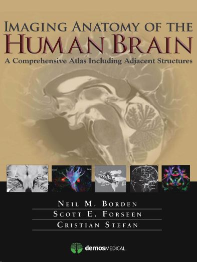
This volume presents a detailed and beautifully illustrated tour of neuroanatomy, employing…standard and advanced modalities… Ample use of color illustrates fiber tracts and differentiates different nuclei and other structures. The sections are amply labeled and easy to follow, allowing the reader to become familiar with cognate areas on images of multiple techniques. — Carl E. Stafstrom, Division of Pediatric Neurology, Johns Hopkins Hospital, Journal of Pediatric Epilepsy
An Atlas for the 21st Century
Key Features:
- Provides detailed views of anatomic structures within and around the human brain utilizing over 1,000 high quality images across a broad range of imaging modalities
- Contains extensively labeled images of all regions of the brain and adjacent areas that can be compared and contrasted across modalities
- Includes specially created color illustrations using computer 3-D modeling techniques to aid in identifying structures and understanding relationships
- Goes beyond a typical brain atlas with detailed imaging of skull base, calvaria, facial skeleton, temporal bones, paranasal sinuses, and orbits
- Serves as an authoritative learning tool for students and trainees and practical reference for clinicians in multiple specialties


